free counters

الاثنين، 24 أغسطس 2009

Encyclopedia of Global Health


"Encyclopedia of Global Health" ed. by Yawei Zhang
Sage Publications, Inc | 2008 | ISBN: 1412941865 | 2046 pages | PDF | 23 Mb 


A general reference for topics related to health worlwide, this encyclopedia is ambitious in its scope, with entries for specific diseases and conditions, geographical areas, health issues, biographical information, and organizations related to world health policy. This book includes 1200 entries from "Abortion" to "Zoll. An alphabetical contents list is included in each volume. The thorough entries vary in length, and each one concludes with a bibliography and cross-references
download link

An Anatomy of Thought: The Origin and Machinery of the Mind


An Anatomy of Thought: The Origin and Machinery of the Mind 
Posted By : Book-er 

Ian Glynn "An Anatomy of Thought: The Origin and Machinery of the Mind"
Oxford University Press | English | 2003-04-10 | ISBN: 0195158032 | 464 pages | PDF | 25 MB


Drawing on a dazzling array of disciplines--physiology, neurology, psychology, anthropology, linguistics, and philosophy--Ian Glynn explains virtually every aspect of the workings of the brain, unlocking the mysteries of the mind. Glynn writes with exceptional clarity as he illuminates the mechanics of nerve messages; the functioning of sensory receptors; the processes by which the brain sees, tastes, and smells; the seats of language, memory, and emotions. The breadth of Glynn's erudition is astonishing, as he ranges from parallel processing in computers to the specialization of different regions of the brain (illustrated with fascinating instances of the bizarre effects of localized brain damage). He explains the different types of memory, traces the path of information that leads to emotional responses, and engages in a discussion of language that ranges from Noam Chomsky to Hawaiian pidgin. No other single volume has captured the full expanse of our knowledge of consciousness and the brain. A work of unequaled authority and eloquence, this book promises to be a new landmark of scientific writing. "Monumental."--The Observer "Glynn's erudition is astonishing...a hugely enjoyable intellectual journey."--Nature
To start download free click HERE:
 Download many interesting free eBooks HERE

الجمعة، 21 أغسطس 2009

Drug-Induced Hepatotoxicity


Drugs are an important cause of liver injury. More than 900 drugs, toxins, and herbs have been reported to cause liver injury, and drugs account for 20-40% of all instances of fulminant hepatic failure. Approximately 75% of the idiosyncratic drug reactions result in liver transplantation or death. Drug-induced hepatic injury is the most common reason cited for withdrawal of an approved drug. Physicians must be vigilant in identifying drug-related liver injury because early detection can decrease the severity of hepatotoxicity if the drug is discontinued. The manifestations of drug-induced hepatotoxicity are highly variable, ranging from asymptomatic elevation of liver enzymes to fulminant hepatic failure. Knowledge of the commonly implicated agents and a high index of suspicion are essential in diagnosis.

For excellent patient education resources, visit eMedicine's Poisoning Center, Public Health Center, and Substance Abuse Center. Also, see eMedicine's patient education articles Acetaminophen (Tylenol) Poisoning, FDA Overview, Pain Medications, and Alcoholism.

Mortality/morbidity

In the United States, approximately 2000 cases of acute liver failure occur annually and drugs account for over 50% of them (39% are due to acetaminophen, 13% are idiosyncratic reactions due to other medications). Drugs account for 2-5% of cases of patients hospitalized with jaundice and approximately 10% of all cases of acute hepatitis.

Internationally, data on the incidence of adverse hepatic drug reactions in the general population remain unknown.

Drugs withdrawn from the market secondary to hepatotoxicity

In the last few years, the US Food and Drug Administration (FDA) has withdrawn 2 drugs from the market for causing severe liver injury: bromfenac and troglitazone. Bromfenac (Duract), a nonsteroidal anti-inflammatory drug (NSAID), was introduced in 1997 as a short-term analgesic for orthopedic patients. Although approved for a dosing period of less than 10 days, patients used it for longer periods. This resulted in more than 50 cases of severe hepatic injury, and the drug had to be withdrawn in 1998. Troglitazone (Rezulin) is a thiazolidinedione and was approved in 1997 as an antidiabetic agent. Over 3 years, more than 90 cases of hepatotoxicity were reported, which resulted in withdrawal of this drug.

Kava kava, an herb used for anxiety, was reported as being hepatotoxic and was withdrawn from the German market.1 The FDA has also issued a warning in this country. This demonstrates the importance of postmarketing surveillance to identify reactions that are not reported or are underreported in drug trials.

Pemoline (Cylert), used for attention deficit disorder and narcolepsy is no longer available in the United States. The Food and Drug Administration (FDA) concluded that the overall risk of liver toxicity from pemoline outweighs the benefits. In May 2005, Abbott chose to stop sales and marketing of their brand of pemoline (Cylert) in the U.S. In October 2005, all companies that produced generic versions of pemoline also agreed to stop sales and marketing of pemoline.

Other drugs that have significant limitations of use because of their hepatotoxic effects are felbamate (Felbatol), an antiepileptic used for complex partial seizures; zileuton (Zyflo), indicated for asthma; tolcapone (Tasmar), used for Parkinson disease; trovafloxacin (Trovan), an antibiotic; benoxaprofen, an NSAID; and tienilic acid, a diuretic.

Recent warnings issued by the FDA
In June 2009, the US Food and Drug Administration (FDA) issued a report that identified 32 cases (22 adult and 10 pediatric) of serious liver injury associated with propylthiouracil (PTU). Of the adults, 12 deaths and 5 liver transplants occurred, and among the pediatric patients, 1 death and 6 liver transplants occurred. PTU is indicated for hyperthyroidism due to Graves disease.

These reports suggest an increased risk for liver toxicity with PTU compared with methimazole. Serious liver injury has been identified with methimazole in 5 cases (3 resulting in death). PTU is considered as second-line drug therapy, except in patients who are allergic or intolerant to methimazole, or for women who are in the first trimester of pregnancy. Rare cases of embryopathy, including aplasia cutis, have been reported with methimazole during pregnancy.

The FDA recommends the following criteria be considered for prescribing PTU. For more information see the FDA Safety Alert.
Reserve PTU use during first trimester of pregnancy, or in patients who are allergic to or intolerant of methimazole.
Closely monitor PTU therapy for signs and symptoms of liver injury, especially during the first 6 months after initiation of therapy.
For suspected liver injury, promptly discontinue PTU therapy and evaluate for evidence of liver injury and provide supportive care.
PTU should not be used in pediatric patients unless the patient is allergic to or intolerant of methimazole, and no other treatment options are available.
Counsel patients to promptly contact their health care provider for the following signs or symptoms: fatigue, weakness, vague abdominal pain, loss of appetite, itching, easy bruising, or yellowing of the eyes or skin.

Severe hepatic injury, including cases of hepatic failure, has been reported in patients taking interferon beta-1a (Avonex) used in treatment of multiple sclerosis. Asymptomatic elevation of hepatic transaminases have also been reported and, in some patients, recurred upon rechallenge. In some cases, these events occurred in the presence of other drugs that have been associated with hepatic injury. The potential risk of Avonex used in combination with known hepatotoxic drugs or other products (eg, alcohol) should be considered prior to Avonex administration or when adding new agents to the regimen of patients already on Avonex.

In January 2006, the US Food and Drug Administration (FDA) issued a warning after 3 cases of serious liver toxicity were reported with taking telithromycin. In June 2006, the prescribing information for telithromycin (Ketek) was changed to include a warning describing the drug's association with rare cases of serious liver injury and liver failure. Four of these events resulted in deaths and one resulted in liver transplant. The added warning follows evaluation by the FDA on postmarketing surveillance reports. If clinical hepatitis or liver enzyme elevations combined with other systemic symptoms occur, telithromycin should be permanently discontinued. Telithromycin is an antibiotic of the ketolide class, approved by the FDA in April 2004 for the treatment of respiratory infections in adults. It is marketed and is widely used in several countries including Japan and countries in Europe.

In February 2007, the FDA took further action and removed 2 of the 3 indications: acute bacterial sinusitis and acute bacterial exacerbations of chronic bronchitis. Following comprehensive scientific analysis, the FDA determined that the balance of benefits and risks no longer supports the approval of the drug for these indications. Telithromycin is now indicated for treatment of mild-to-moderate community-acquired pneumonia.

In October 2005, the manufactures of duloxetine (an anti-depressant) reported postmarketing cases of hepatitis and cholestatic jaundice. The new package insert now states, "Cymbalta should not be administered to patients with substantial alcohol use or any hepatic insufficiency."

Risk factors for drug-induced liver injury 
Race: Some drugs appear to have different toxicities based on race. For example, blacks and Hispanics may be more susceptible to isoniazid (INH) toxicity. The rate of metabolism is under the control of P-450 enzymes and can vary from individual to individual.
Age: Apart from accidental exposure, hepatic drug reactions are rare in children. Elderly persons are at increased risk of hepatic injury because of decreased clearance, drug-to-drug interactions, reduced hepatic blood flow, variation in drug binding, and lower hepatic volume. In addition, poor diet, infections, and multiple hospitalizations are important reasons for drug-induced hepatotoxicity.
Sex: Although the reasons are unknown, hepatic drug reactions are more common in females.
Alcohol ingestion: Alcoholic persons are susceptible to drug toxicity because alcohol induces liver injury and cirrhotic changes that alter drug metabolism. Alcohol causes depletion of glutathione (hepatoprotective) stores that make the person more susceptible to toxicity by drugs.
Liver disease: In general, patients with chronic liver disease are not uniformly at increased risk of hepatic injury. Although the total cytochrome P-450 is reduced, some may be affected more than others. The modification of doses in persons with liver disease should be based on the knowledge of the specific enzyme involved in the metabolism. Patients with HIV infection who are co-infected with hepatitis B or C virus are at increased risk for hepatotoxic effects when treated with antiretroviral therapy. Similarly, patients with cirrhosis are at increased risk of decompensation by toxic drugs.
Genetic factors: A unique gene encodes each P-450 protein. Genetic differences in the P-450 enzymes can result in abnormal reactions to drugs, including idiosyncratic reactions. Debrisoquine is an antiarrhythmic drug that undergoes poor metabolism because of abnormal expression of P-450-II-D6. This can be identified by polymerase chain reaction amplification of mutant genes. This has led to the possibility of future detection of persons who can have abnormal reactions to a drug.
Other comorbidities: Persons with AIDS, persons who are malnourished, and persons who are fasting may be susceptible to drug reactions because of low glutathione stores.
Drug formulation: Long-acting drugs may cause more injury than shorter-acting drugs.
Host factors that may enhance susceptibility to drugs, possibly inducing liver disease
Female - Halothane, nitrofurantoin, sulindac
Male - Amoxicillin-clavulanic acid (Augmentin)
Old age - Acetaminophen, halothane, INH, amoxicillin-clavulanic acid
Young age - Salicylates, valproic acid
Fasting or malnutrition - Acetaminophen
Large body mass index/obesity - Halothane
Diabetes mellitus - Methotrexate, niacin
Renal failure - Tetracycline, allopurinol
AIDS - Dapsone, trimethoprim-sulfamethoxazole
Hepatitis C - Ibuprofen, ritonavir, flutamide
Preexisting liver disease - Niacin, tetracycline, methotrexate

Pathophysiology and mechanisms of drug-induced liver injury 
Pathophysiologic mechanisms: The pathophysiologic mechanisms of hepatotoxicity are still being explored and include both hepatocellular and extracellular mechanisms. The following are some of the mechanisms that have been described:
Disruption of the hepatocyte: Covalent binding of the drug to intracellular proteins can cause a decrease in ATP levels, leading to actin disruption. Disassembly of actin fibrils at the surface of the hepatocyte causes blebs and rupture of the membrane.
Disruption of the transport proteins: Drugs that affect transport proteins at the canalicular membrane can interrupt bile flow. Loss of villous processes and interruption of transport pumps such as multidrug resistance–associated protein 3 prevent the excretion of bilirubin, causing cholestasis.
Cytolytic T-cell activation: Covalent binding of a drug to the P-450 enzyme acts as an immunogen, activating T cells and cytokines and stimulating a multifaceted immune response.
Apoptosis of hepatocytes: Activation of the apoptotic pathways by the tumor necrosis factor-alpha receptor of Fas may trigger the cascade of intercellular caspases, which results in programmed cell death.
Mitochondrial disruption: Certain drugs inhibit mitochondrial function by a dual effect on both beta-oxidation energy production by inhibiting the synthesis of nicotinamide adenine dinucleotide and flavin adenine dinucleotide, resulting in decreased ATP production.
Bile duct injury: Toxic metabolites excreted in bile may cause injury to the bile duct epithelium.
Drug toxicity mechanisms: The classic division of drug reactions is into at least 2 major groups, (1) drugs that directly affect the liver and (2) drugs that mediate an immune response.
Intrinsic or predictable drug reactions: Drugs that fall into this category cause reproducible injuries in animals, and the injury is dose related. The injury can be due to the drug itself or to a metabolite. Acetaminophen is a classic example of a known intrinsic or predictable hepatotoxin at supertherapeutic doses. Another classic example is carbon tetrachloride.
Idiosyncratic drug reactions: Idiosyncratic drug reactions can be subdivided into those that are classified as hypersensitivity or immunoallergic and those that are metabolic-idiosyncratic.
Hypersensitivity: Phenytoin is a classic, if not common, cause of hypersensitivity reactions. The response is characterized by fever, rash, and eosinophilia and is an immune-related response with a typical short latency period of 1-4 weeks.
Metabolic-idiosyncratic: This type of reaction occurs through an indirect metabolite of the offending drug. Unlike intrinsic hepatotoxins, the response rate is variable and can occur within a week or up to one year later. It occurs in a minority of patients taking the drug, and no clinical manifestations of hypersensitivity are noted. INH toxicity is considered to fall into this class. Not all drugs fall neatly into one of these categories, and overlapping mechanisms may occur with some drugs (eg, halothane).
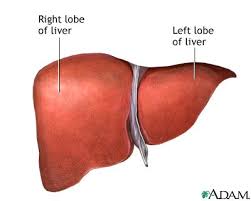
Anatomy of The Liver
The Couinaud classification of liver anatomy divides the liver into eight functionally indepedent segments. 
Each segment has its own vascular inflow, outflow and biliary drainage.
In the centre of each segment there is a branch of the portal vein, hepatic artery and bile duct.
In the periphery of each segment there is vascular outflow through the hepatic veins.

Right hepatic vein divides the right lobe into anterior and posterior segments.
Middle hepatic vein divides the liver into right and left lobes (or right and left hemiliver). This plane runs from the inferior vena cava to the gallbladder fossa.
Left hepatic vein divides the left lobe into a medial and lateral part.

Portal vein divides the liver into upper and lower segments.
The left and right portal veins branch superiorly and inferiorly to project into the center of each segment.

Because of this division into self-contained units, each segment can be resected without damaging those remaining. For the liver to remain viable, resections must proceed along the vessels that define the peripheries of these segments. This means, that resection-lines parallel the hepatic veins,
The centrally located portal veins, bile ducts, and hepatic arteries are preserved.
 

Clockwise numbering of the segments Segments numbering

There are eight liver segments.
Segment 4 is sometimes divided into segment 4a and 4b according to Bismuth.
The numbering of the segments is in a clockwise manner (figure).
Segment 1 (caudate lobe) is located posteriorly. It is not visible on a frontal view.
 

On a frontal view of the liver the posteriorly located segments 6 and 7 are not visible. 
The illustrations above are schematic presentations of the liversegments. 
In reality however the proportions are different.
On a normal frontal view the segments 6 and 7 are not visible because they are located more posteriorly. 
The right border of the liver is formed by segment 5 and 8.
Although segment 4 is part of the left hemiliver, it is situated more to the right.

Couinaud divided the liver into a functional left and right liver (in French 'gauche et droite foie') by a main portal scissurae containing the middle hepatic vein. This is known as Cantlie's line. 
Cantlie's line runs from the middle of the gallbladder fossa anteriorly to the inferior vena cava posteriorly.

On this illustration it looks as if the medial part of the left lobe is separated from the lateral part by the falciform ligament. However it actually is the left hepatic vein, that separates the medial part (segment 4) from the lateral part (segments 2 and 3).
The left hepatic vein is located slightly to the left of the falciform ligament.
 

LEFT: above the level of the left portal vein.
RIGHT: at the level of the left portal vein Transverse anatomy

The far left figure is a transverse image through the superior liver segments, that are divided by the hepatic veins.

The right figure shows a transverse image at the level of the left portal vein.
At this level the left portal vein divides the left lobe of the liver into the superior segments (2 and 4A) and the inferior segments (3 and 4B).
The left portal vein is at a higher level than the right portal vein.
 

LEFT: at the level of the right portal vein.
RIGHT: at the level of the splenic vein. 
The image on the far left is at the level of the right portal vein. At this level the right portal vein divides the right lobe of the liver into superior segments (7 and 8) and the inferior segments (5 and 6).
The level of the right portal vein is inferior to the level of the left portal vein.

At the level of the splenic vein, which is below the level of the right portal vein, only the inferior segments are seen (right image).
 

Hypertrophy of caudate lobe in a patient with livercirrhosis. Notice the small lobulated right hemiliver. Caudate lobe

The caudate lobe or segment 1 is located posteriorly. 
The caudate lobe is anatomically different from other lobes in that it often has direct connections to the IVC through hepatic veins, that are separate from the main hepatic veins.
The caudate lobe may be supplied by both right and left branches of the portal vein.
On the left a patient with cirrhosis with extreme atrophy of the right lobe, normal volume of the left lobe and hypertrophy of the caudate lobe.
Due to a different blood supply the caudate lobe is spared from the disease process and hypertrophied to compensate for the loss of normal liverparenchyma


Neurovascular Neuropsychology


Neurovascular Neuropsychology
Springer | ISBN: 90387707131 | 22009-05-15 | PDF | 566 pages | 4 Mb

Download Link

Cosmetic Dermatology : Principles and Practice


Cosmetic Dermatology : Principles and Practice, 2nd Edition
McGraw-Hill Professional | 2009-04-08 | ISBN: 0071490620 | 336 Pages | PDF | 17 MB


Download Link

Anatomy of the Human Body Henry Gray


Anatomy of the Human Body
 
Henry Gray
 
The Bartleby.com edition of Gray’s Anatomy of the Human Body features 1,247 vibrant engravings—many in color—from the classic 1918 publication, as well as a subject index with 13,000 entries ranging from the Antrum of Highmore to the Zonule of Zinn
TWENTIETH EDITION
THOROUGHLY REVISED AND RE-EDITED BY WARREN H. LEWIS
ILLUSTRATED WITH 1247 ENGRAVINGS

PHILADELPHIA: LEA & FEBIGER, 1918
NEW YORK: BARTLEBY.COM, 2000
I. Embryology
The Extremities
http://www.bartleby.com/107/49.html
http://www.bartleby.com/107/50.html
http://www.bartleby.com/107/51.html
http://www.bartleby.com/107/52.html
http://www.bartleby.com/107/54.html
http://www.bartleby.com/107/55.htm 

http://www.bartleby.com/107/57.html
http://www.bartleby.com/107/61.html
vessels
http://www.bartleby.com/107/142.html
http://www.bartleby.com/107/157.html
http://www.bartleby.com/107/165.html

Stem Cells , Human Embryos and Ethics : Interdisciplinary Perspectives


Stem Cells , Human Embryos and Ethics : Interdisciplinary Perspectives 
Publisher: Springer | ISBN: 140206988X | Edition 2008 | PDF | 268 Pages | 2,2 MB


Download Link

الجمعة، 14 أغسطس 2009

Forensic Science


Forensic Science
Salem Press | 2008-10-17 | ISBN : 1587654239 | Pages: 1056 | PDF | 17.33 MB


Download Link


Recent Advances in Transthyretin Evolution, Structure and Biological Functions


Recent Advances in Transthyretin Evolution , Structure and Biological Functions
Springer | 2009 | ISBN: 3642006450 | 374 Pages | PDF | 5 MB


Download Link
0R


الأربعاء، 12 أغسطس 2009

Alzheimer’s Disease Symptoms and Stages


Alzheimer’s disease is the most common form of dementia, a serious brain disorder that impacts daily living through memory loss and cognitive changes. Alzheimer’s is a degenerative disease, progressing from mild forgetfulness to widespread neurological impairment and ultimately death. Chemical and structural changes in the brain gradually destroy the ability to create, remember, learn, reason, and relate to others. As critical cells die, drastic personality loss occurs and body systems fail.
Although not all memory loss indicates Alzheimer’s disease, one in ten people over 65 years of age, and over half of those 85+ have Alzheimer’s disease. Currently, 26 million people worldwide have this dementia, and over 15 million Americans will be affected by the year 2050.
Early-onset Alzheimer’s effects patients under the age of 65. This relatively rare condition is seen more often when parents or grandparents developed Alzheimer’s disease at a young age, and is generally associated with three genetic markers.

Signs and symptoms that may mean Alzheimer’s disease

For many, recognition of memory problems in themselves or a loved one brings fear of Alzheimer’s disease. Age-related brain shrinkage does produce normal changes in processing speed, attention, and short term memory, creating so-called “senior moments.” Understanding significance begins with knowing what is normal.SIGNS OF NORMAL CHANGE VS. EARLY ALZHEIMER’S SYMPTOMS

*Normal
$Early Alzheimer’s disease

*Can’t find your keys 
$Routinely place important items in odd places

*Search for casual names and words 
$Forget names of family and common objects

*Briefly forget conversation details 
$Frequently forget entire conversations

*Can’t find a recipe 
$Can’t follow recipe directions

*Forget to write down a check 
$Can no longer manage checkbook

*Cancel a date with friends 
$Withdraw from usual interests

*Miss an occasional right turn 
$Get lost in familiar places

What else might be causing these symptoms?

Significant cognitive and memory losses are not symptoms of normal aging. Most people over 65 experience a level of forgetfulness that is merely inconvenient and generally involves unimportant information. Terms like eccentric and senile can mask fears of Alzheimer’s disease, while other conditions may actually be causing mental decline.  

Symptoms that mimic early Alzheimer’s disease may result from:
Central nervous system and other degenerative disorders-, head injuries, brain tumors, stroke, epilepsy, Pick’s Disease, Parkinson’s disease, Huntington’s disease 
Metabolic ailments- hypothyroidism, hypoglycemia, malnutrition, vitamin deficiencies, dehydration, kidney or liver failure
Substance-induced conditions-drug interactions, medication side-effects, alcohol and drug abuse
Psychological factors- dementia syndrome, depression, emotional trauma, chronic stress, psychosis, chronic sleep deprivation, delirium 
Infections- meningitis, encephalitis, and syphilis
What to do if you notice Alzheimer’s symptoms

Research shows Alzheimer’s disease causes more worry than any other condition for Americans over 55 years of age. If your family history includes Alzheimer’s and you find yourself forgetting things, it doesn’t necessarily mean you have this disease. Even when you fear the worst, it is important to share your concerns and seek expert advice.  
Why not just wait and see?  

When symptoms appear suddenly, it is critical to seek medical attention. Conditions such as stroke, drug interactions, tumors, and seizures should be treated immediately. Timely intervention may also control or eliminate symptoms from other physical and psychological factors. 

If your symptoms are the result of Alzheimer’s disease, medications can delay the onset of more debilitating symptoms. Early diagnosis can prolong independence and is the first step towards treatment, management, and living life fully.
Ten Warning Signs of Alzheimer’s 

According to the Alzheimer’s Association, the ten warning signs are:
Memory loss
Difficulty performing familiar tasks
Problems with language
Disorientation to time and place
Poor or decreased judgment
Problems with abstract thinking
Misplacing things
Changes in mood or behavior
Changes in personality
Loss of initiative
Where should I go if I suspect Alzheimer’s?

Although no single test for Alzheimer’s exists, a skilled general practitioner, neurologist, or geriatrician can make a reasonable diagnosis by excluding other causes of your symptoms. Begin by preparing a list of concerns to discuss with your doctor. Ask a family member or friend to go with you to take notes and help you remember your questions. If your doctor suspects Alzheimer’s disease and recommends further assessment, ask for support and information from your local Alzheimer’s Association while you wait for a diagnosis.
Can I prevent or inhibit Alzheimer’s disease?

With each forgotten name or misplaced item, memory loss can be frightening. Maintaining a brain-healthy lifestyle, understanding controllable risk factors, and participating in research studies, can increase your sense of empowerment and protect your memory. 

Incest


Incest refers to any sexual activity between closely related persons (often within the immediate family) that is illegal or socially taboo. The type of sexual activity and the nature of the relationship between persons that constitutes a breach of law or social taboo vary with culture and jurisdiction. Some societies consider it to include only those who live in the same household, or who belong to the same clan or lineage; other societies consider it to include "blood relatives"; other societies further include those related by adoption or marriage.[1]

According to some studies,[2] the most frequently reported type of incest is father-daughter incest. However, others studies[3][4][5] suggest that sibling incest occurs as often, or more often, than other types of incest. Incest between adults and prepubescent or adolescent children is considered a form of child sexual abuse[6][7] that has been shown to be one of the most extreme forms of childhood trauma, a trauma that often does serious and long-term psychological damage, especially in the case of parental incest.[8] Prevalence is difficult to generalize, but research has estimated 10-15% of the general population as having at least one incest experience, with less than 2% involving intercourse or attempted intercourse.[9] Among women, research has yielded estimates as high as twenty percent.[8]

Consensual adult incest is rarely reported.[citation needed] Consensual incest between adults is criminalized in most countries,[10] although it is seen by some as a victimless crime.[11][12]

Most societies have some form of incest avoidance.[13][14] The incest taboo is and has been one of the most common of all cultural taboos, both in current nations and many past societies,[15] with legal penalties imposed in some jurisdictions. Most modern societies have legal or social restrictions on closely consanguineous marriages.[16] However, in some societies, such as that of Ancient Egypt, brother–sister, father–daughter, and mother–son relations were practiced among royalty.[17][18] In addition, the Balinese[19] and some Inuit tribes[20] have altogether different beliefs about what constitutes illegal and immoral incest.

الاثنين، 10 أغسطس 2009

Acland’s DVD Atlas of Human Anatomy

Acland’s DVD Atlas of Human Anatomy
مع شرح واضح ومميز بصوت الدكتور
Robert Acland
download links


The Upper Extremity: Disc 1

http://mihd.net/c5uk3t

http://mihd.net/r6mot5

http://mihd.net/gvmh45

http://mihd.net/yknt3r

http://mihd.net/nwdog4

http://mihd.net/0hxm9q

http://mihd.net/3niov8


(IMG:http://bramjnet.medical.googlepages.com/spact24.gif) 


The Lower Extremity: Disc 2


http://mihd.net/f7b5t9

http://mihd.net/9yi1l6

http://mihd.net/bworu1

http://mihd.net/cv1slp

http://mihd.net/6arhku

http://mihd.net/jxhv8f

http://mihd.net/laxirq


(IMG:http://bramjnet.medical.googlepages.com/spact24.gif) 


Trunk : Disc 3


http://mihd.net/wt3pq6

http://mihd.net/t4mrcb

http://mihd.net/l96oin

http://mihd.net/5dtqry

http://mihd.net/dsoxb5

http://mihd.net/ovhjl6

http://mihd.net/m0zvac


(IMG:http://bramjnet.medical.googlepages.com/spact24.gif) 


The Head And Neck, Part 1, Disc 4


http://mihd.net/d9wtiv

http://mihd.net/hntywj

http://mihd.net/dl6vio

http://mihd.net/dqfjm2

http://mihd.net/zxehpt

http://mihd.net/f54nk3

http://mihd.net/cd9v71


(IMG:http://bramjnet.medical.googlepages.com/spact24.gif) 


The Head And Neck, Part 2, Disc 5


http://mihd.net/bfc2ha

http://mihd.net/agr7kj

http://mihd.net/hla2wk

http://mihd.net/jt25pz

http://mihd.net/y5ordn

http://mihd.net/jroq0z

http://mihd.net/o07y9x


(IMG:http://bramjnet.medical.googlepages.com/spact24.gif) 


The Internal Organs: Disc 6


http://mihd.net/03nouw

http://mihd.net/tgyhd5

http://mihd.net/z4puec

http://mihd.net/05f4dh

http://mihd.net/t06g1l

http://mihd.net/xmzvlo

http://mihd.net/9gx0oz


كلمة المرور لجميع الأجزاء السابقة :-

Password: xpmisho-dvd4arab

European Heart Journal - April 2009 (Vol.30 - N°7)

European Heart Journal - April 2009 (Vol.30 - N°7)
English | 135 pages | PDF | 4.88 MB
download link
or

German : A Linguistic Introduction


German : A Linguistic Introduction
Cambridge University Press | 2009 | ISBN: 0521852854, 0521618037 | 332 Pages | PDF | 1,2 MB

download link


Or


الأحد، 9 أغسطس 2009

Too Much Iron: Iron Overdose


Iron is a mineral that functions primarily as a carrier of oxygen in the body, both as a part of hemoglobin in the blood and of myoglobin in the muscles. The body increases or decreases iron absorption according to need. The presence of vitamin C (ascorbic acid) in a meal increases iron absorption. The body absorbs iron more efficiently when iron stores are low, and during growth spurts or pregnancy.

The most common indication of poor iron status is iron deficiency anemia, a condition in which the size and number of red blood cells are reduced. This condition may result from inadequate intake of iron or from blood loss. 
When there is insufficient iron from dietary sources, or as a result of blood loss in the body, the amount of hemoglobin in the bloodstream is reduced and oxygen cannot be efficiently transported to tissues and organs throughout the body. Iron-deficiency anemia is characterized by fatigue, shortness of breath, pale skin, concentration problems, dizziness, a weakened immune system, and energy loss.

Inadequate intake or iron can cause ill-health but just as important as a cause of illness is consuming too much of Iron. Indeed, Iron excess is a greater risk than iron deficiency for many older Americans. In a study of more than 1,000 white men and women aged 67 to 96 who live at home, 13 percent had too much iron in their blood, but only three percent had too little. 

Healthy people usually absorb about 10 percent of the iron contained in the food they eat to meet the body needs. This is why we need to take iron supplements. But wait a minute. We need to get a few things clear. Let's define anemia: a deficiency of red cells or hemoglobin, or red cells that die too young or are discolored or possess an abnormal shape, or red cells that lack adequate iron. Now defining iron deficiency vary from lab to lab. Most "normal" levels are set too high. Saturation: 12 to 40-45% is reasonable at the present time. Ferritin: 5 to probably 50. Think about it. If "normal" levels are set artificially high, and your levels fall below that "normal," you are "iron deficient."

So how much iron do you really need? Iron is not excreted. The iron you absorb stays and accumulates in storage except that you can lose one milligram a day through hair, finger nails, skin cells and other detritus. That is the amount needed every day to replace the loss. One milligram, that’s all and for women in reproductive years, one and a half milligram. The other way to lose iron, of course, is by blood loss.

The other thing to note is that hemoglobin is not iron! Yes, you are anemic if your hemoglobin is low but that does not necessarily mean your iron is low. Indeed, what might be happening is that the iron is collecting in storage instead of going into hemoglobin. You are actually iron-loaded and need iron removed despite the anemia. The anemia should be treated with B vitamins, especially B12, B6 and folic acid. Many patients with anemia are dying of iron overload, and some are hastened to their death by their physicians who give them more iron.

Even a small amount of excess iron can damage heart and brain and other storage sites in the body and lead to heart attack or stroke. There is exaggerated concern when hemoglobin falls temporarily, following surgery, for example. Blood transfusions are over-used. A study shows that surgery patients who do not receive transfusions survive better than those who do.1 

Before taking iron you must test saturation and ferritin. Ferritin indicates storage iron, which is not essential to maintain life. If both saturation and ferritin are extremely low, you must find out why. Low iron is a signal that iron is being used by cancer cells or is feeding bacteria, or usually it means there is chronic daily blood loss. The bleeding could be from an ulcer or tumor, etc. The source must be found.

Iron is in just about everything. If you are not absorbing the one daily milligram, you are truly on a starvation diet, and low iron is the least of your worries. So, go for iron supplementation only when you need it and be aware of iron's toxic ability to harm you. 

Severe iron overload, which causes liver and heart damage, can occur in people who are genetically susceptible. This is called haemochromatosis. Haemochromatosis, also called iron storage disease, or bronze diabetes is an inborn metabolic defect characterized by an increased absorption of iron, which accumulates in body tissues. The body has no natural way to rid itself of excess iron, so extra iron is stored in body tissues, especially the liver, heart, and pancreas.

People with haemochromatosis absorb more than the body needs The clinical manifestations include skin pigmentation, diabetes, enlargement of the spleen and liver, heart failure, and general weakness and lassitude. In males, the symptoms are usually noted after 35 years of age, and in females, after menopause, when iron is no longer lost through menstruation and pregnancy. Treatment consists of the removal of blood at regular intervals to decrease the concentration of body iron.

Excess stored iron can lead to atherosclerosis and ischemic heart disease. Phlebotomy, or blood removal, has been used to reduce stored iron in patients with iron overload with some success. Iron chelation with drugs such as desferrioxamine (Desferal) that help patients excrete excess stores of iron can be helpful in treating iron overload caused by multiple blood transfusions.

Plague


There were 75 million people that were killed by the Black Death, also known as the Bubonic There were 75 million people that were killed by the Black Death, also known as the Bubonic Plague. The Black Death was in reference to one of three related diseases, one of which was the Bubonic Plague. There is no real evidence of the total body count. They are educated guesses, only that, conjecture. Though rough estimates, they are probably still close enough to inform people.

It wiped out roughly 1/3 of Europe's population, which means it killed about 25-50 million just there.


Estimates put the death toll of the Black Death or plague around 25 Million, or 25-50% of the population of Europe. The plague was first noted in 1347 and carried on for about 3 or 4 years until 1350/51. It is hard to say exactly how many were killed per year, but based on the above information I would estimate about 8.33 Million died each year during the plagues prominence in Europe.
. The Black Death was in reference to one of three related diseases, one of which was the Bubonic Plague. There is no real evidence of the total body count. They are educated guesses, only that, conjecture. Though rough estimates, they are probably still close enough to inform people. 

It wiped out roughly 1/3 of Europe's population, which means it killed about 25-50 million just there 

Estimates put the death toll of the Black Death or plague around 25 Million, or 25-50% of the population of Europe. The plague was first noted in 1347 and carried on for about 3 or 4 years until 1350/51. It is hard to say exactly how many were killed per year, but based on the above information I would estimate about 8.33 Million died each year during the plagues prominence in Europe.

Atlas of the Human Brain Stem and Cerebellum


Thomas P. Naidich, Henri M. Duvernoy, Bradley N. Delman, A. Gregory Sorensen "Duvernoy's Atlas of the Human Brain Stem and Cerebellum: High-Field MRI, Surface Anatomy, Internal Structure, Vascularization and 3 D Sectional Anatomy"
Springer | English | 2008-12-12 | ISBN: 321173970X | 876 pages | PDF | 154 MB


download link
or

TeLinde's Operative Gynecology


John A Rock, Howard W Jones, "TeLinde's Operative Gynecology" 
Lippincott Williams & Wilkins; Tenth Edition (April 1, 2008) | ISBN-10: 0781772346 | 1472 Pages | CHM | 141,3 MB

download link
or

Basic Concepts of Molecular Pathology

Basic Concepts of Molecular Pathology (Molecular Pathology Library)
Springer | ISBN: 0387896252 | 2009-06-16 | PDF | 224 Pages | 3 Mb

download link
or

Magnetism in Medicine


Magnetism in Medicine 
Publisher: Wiley-VCH | ISBN: 3527405585 | Edition 2007 | PDF | 655 Pages | 16,26 MB

download link
or